Applications / Microscopy and inspection
Depth and extended focus from a single snapshot
A microscope objective sees only a thin slice of the sample sharply, and its blur looks the same above focus and below. A phase mask at the aperture, with matched software, changes what one snapshot holds: the height of a surface, the depth of a defect, or an image that stays sharp through several times the usual depth of field. The aim is to measure and inspect without scanning through focus.
- Status
- Shown on research microscopes, in surface metrology and biological imaging
- Height precision
- About 30 nm over a 4 µm range, on a tilted silicon wafer
- Depth of field
- Extended up to about 3×
Why scanning costs throughput
At high magnification the depth of field is about a micrometer or less. A sample that is tilted, warped, stepped or simply thicker than that has to be refocused or scanned slice by slice, and every slice costs acquisition time, data and stage motion.
Autofocus has the same root. A blurred image does not say which way focus lies, so the instrument has to search for it.
What the mask changes
A phase mask at the aperture plane of the objective reshapes the image of every point in the sample. The shape is designed for the job (Fig. 1).
Measure depth
A depth-encoding mask gives each point a shape that changes steadily through focus. Software reads the shape as a height: for surface profiles, for the depth of a defect or particle, and for volumetric imaging of transparent samples.
Extend focus
An extended-focus mask does the opposite. It keeps the response nearly constant through focus, so that one processed image is sharp over up to about 3× the usual depth of field.
Find focus in one shot
An encoded shape shows both how far a surface is from focus and in which direction. One frame can then do the work of a focus sweep.
Encode color
A diffractive element can also write wavelength into the shape of the response, for spectral imaging through a single channel.4
What has been shown
Surface profiling without scanning
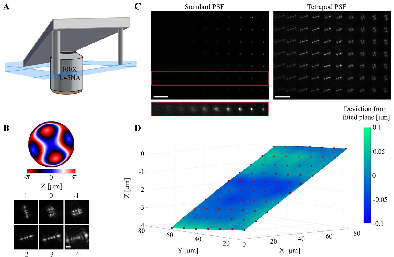
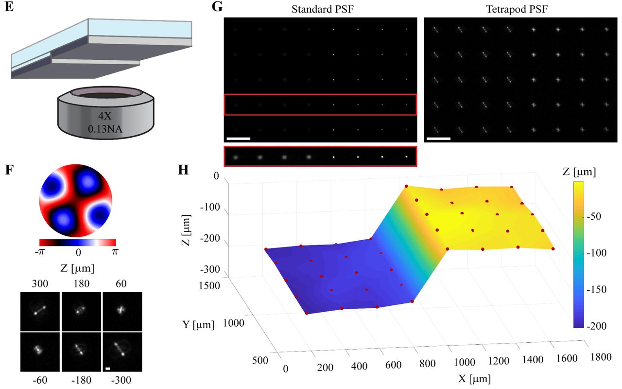
A pattern of light spots is projected onto a reflective surface, and the shape of each spot’s image gives the local height.1 On a silicon wafer tilted under a 100× objective, 70 spots mapped a height range of 4 µm in one frame, with an axial precision of about 30 nm (Fig. 2). With a 4× objective the same method measured a step of about 200 µm across a field more than a millimeter wide (Fig. 3).
Extended depth of field
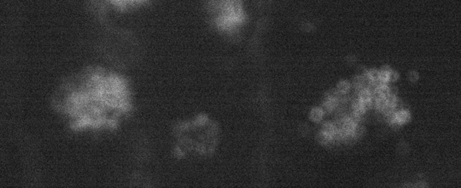
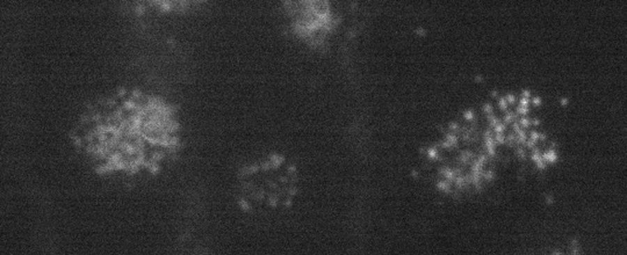
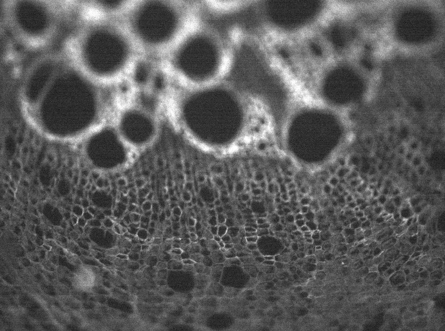
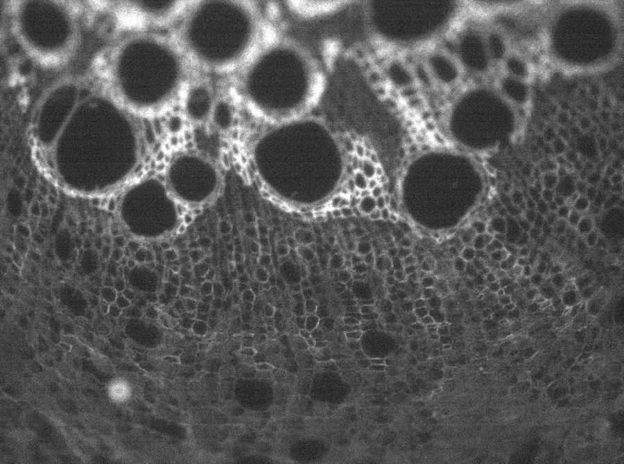
Fig. 4 shows two samples, each imaged with a standard objective and with an extended-focus mask: dots printed on both faces of a transparent sheet 100 µm thick, and a tilted sample, where the depth of field grew by about 1.7×.
Standard
Extended focus
Standard
Extended focus
Dense scenes and single snapshots
Where points sit so close together that their shapes overlap, a neural network reads the positions directly. In single-molecule microscopy this gave a resolution of about 50 nm in all three dimensions.2
In a high-throughput microscope with the mask built into the objective, single snapshots took the place of focus stacks, and acquisition was about 10 times faster.3
What it needs
- A mask is designed for one objective and one wavelength band.
- It has to sit at the aperture plane of the objective, which is reached through a relay at the camera port or inside the objective.
- Height measurement needs something to read: surface texture, particles or projected spots. On a mirror-like wafer the published work projected the spots.
- Extended focus trades some contrast for range. Computation restores it.
- Published demonstrations so far are in surface metrology and biological microscopy. Single-shot autofocus, defect detection and spectral inspection in industrial tools are what we want to build with partners.
Where it helps
The gain is throughput: fewer focus steps for the same information.
- Semiconductor and wafer inspection: surface height and defect depth without focus scanning, and fast 3D screening of large areas ahead of slower high-resolution tools.
- PCB and electronics inspection, where parts stand at different heights.
- Precision manufacturing and quality control.
- Makers of microscopes and inspection tools who want to offer depth, extended focus or single-shot autofocus on existing optics.
Working with us
We are looking for commercial partners who want to build extended depth of field, 3D defect detection, single-shot autofocus, or volumetric and spectral imaging into their inspection tools and microscopes.
The first step is a short, non-confidential call about your optics and what limits your throughput today.
References
- R. Gordon-Soffer et al., “Microscopic scan-free surface profiling over extended axial ranges by point-spread-function engineering,” Science Advances 6, eabc0332 (2020).
- E. Nehme et al., “DeepSTORM3D: dense 3D localization microscopy and PSF design by deep learning,” Nature Methods 17, 734 (2020).
- N. Opatovski et al., “Depth-enhanced high-throughput microscopy by compact PSF engineering,” Nature Communications 15, 4861 (2024).
- R. Orange Kedem et al., “Near index matching enables solid diffractive optical element fabrication via additive manufacturing,” Light: Science & Applications 12, 222 (2023).